Expert Vascular Diagnostic imaging Services
Accurate, Comprehensive Ultrasounds for
Venous and Arterial Health.

At Elevated Vein Solutions, dba EVS Imaging, our mission is to ELEVATE the standard of vascular care by providing accurate, expert diagnostic imaging to support physicians and help restore patients' comfort, confidence, and quality of life.
We are committed to delivering the highest level of patient care and satisfaction, because YOU CENTERED CARE isn't just our motto, It's our promise!
At EVS Imaging, every patient interaction stays local. When you call our office, you speak directly with our Arizona based team- not an outsourced call center or artificial intelligence scheduling service.
Because circulation care deserves real connection- not automation.


Venous Ultrasound Services
Arterial Ultrasound Services
Additional Ultrasound Services

A deep vein thrombosis (DVT), commonly known as a blood clot, occurs when a clot forms within the deep veins-most often in the calf or thigh. Blood clots may be acute (new) or chronic, also known as post-thrombotic changes from a previous clot. A DVT ultrasound evaluation is a non-invasive vascular study used to determine if a blood clot
A deep vein thrombosis (DVT), commonly known as a blood clot, occurs when a clot forms within the deep veins-most often in the calf or thigh. Blood clots may be acute (new) or chronic, also known as post-thrombotic changes from a previous clot. A DVT ultrasound evaluation is a non-invasive vascular study used to determine if a blood clot is present and assess the condition of the veins and blood flow. Early detection is important, as untreated blood clots can lead to damage of the blood vessels, chronic leg symptoms, or potentially life-threatening complications such as a pulmonary embolism (clot traveling to the lungs).

Chronic venous insufficiency/reflux occurs when the valves within the vein(s) become incompetent or damaged and no longer work properly, causing blood to flow backward and pool in the lower extremities instead of returning efficiently to the heart. This condition may lead to symptoms such as leg achiness, pain, heaviness, swelling, varico
Chronic venous insufficiency/reflux occurs when the valves within the vein(s) become incompetent or damaged and no longer work properly, causing blood to flow backward and pool in the lower extremities instead of returning efficiently to the heart. This condition may lead to symptoms such as leg achiness, pain, heaviness, swelling, varicose veins, skin discoloration, cramping, fatigue, or venous ulcers. A venous insufficiency ultrasound evaluation is a specialized, non-invasive vascular study used to assess blood flow within the veins and determine if reflux (backward flow of blood) is present. Ultrasound allows for detailed evaluation of the superficial and deep venous systems, helping identify the source and severity of venous disease. Early diagnosis can help prevent progression of symptoms, skin damage, worsening varicose veins, and chronic wounds while guiding the most appropriate treatment plan.
The inferior vena cava (IVC) and iliac veins are major veins responsible for returning blood from the lower extremities, pelvis, and abdomen back to the heart. An IVC/Iliac vein ultrasound evaluation is a specialized, non-invasive vascular study used to assess blood flow and evaluate for conditions such as blood clots, compression syndrom
The inferior vena cava (IVC) and iliac veins are major veins responsible for returning blood from the lower extremities, pelvis, and abdomen back to the heart. An IVC/Iliac vein ultrasound evaluation is a specialized, non-invasive vascular study used to assess blood flow and evaluate for conditions such as blood clots, compression syndromes like Iliac Vein Compression (May-Thurner Syndrome), narrowing, obstruction, or post-thrombotic changes. This examination helps identify abnormalities that may contribute to leg swelling, pain, heaviness, venous insufficiency, or circulation issues. Early detection through ultrasound can help prevent worsening symptoms and guide appropriate treatment recommendations.
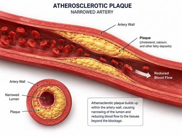
Peripheral arterial disease (PAD) occurs when plaque buildup causes narrowing or blockage within the arteries of the legs, reducing blood flow to the lower extremities. This condition may lead to symptoms such as leg pain or cramping with walking, numbness, weakness, cold feet, discoloration, slow-healing wounds, or pain at rest in more a
Peripheral arterial disease (PAD) occurs when plaque buildup causes narrowing or blockage within the arteries of the legs, reducing blood flow to the lower extremities. This condition may lead to symptoms such as leg pain or cramping with walking, numbness, weakness, cold feet, discoloration, slow-healing wounds, or pain at rest in more advanced cases. A lower extremity arterial duplex ultrasound is a specialized, non-invasive vascular study used to evaluate blood flow within the arteries of the legs and identify areas of narrowing, blockage, or reduced circulation. Ultrasound allows for detailed assessment of arterial waveforms and blood flow velocities, helping determine the severity and location of arterial disease. Early detection of PAD can help prevent worsening circulation problems, tissue damage, non-healing wounds, and reduce the risk of serious cardiovascular complications.
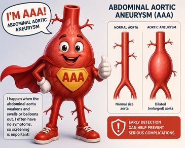
An abdominal aortic aneurysm (AAA) occurs when a weakened area of the abdominal aorta enlarges or balloons outward due to weakening of the artery wall. Over time, the aneurysm may continue to grow and can become life-threatening if rupture occurs. Many aneurysms develop slowly and may not cause symptoms, while others can lead to abdominal
An abdominal aortic aneurysm (AAA) occurs when a weakened area of the abdominal aorta enlarges or balloons outward due to weakening of the artery wall. Over time, the aneurysm may continue to grow and can become life-threatening if rupture occurs. Many aneurysms develop slowly and may not cause symptoms, while others can lead to abdominal, back, or groin pain. An abdominal aortic aneurysm ultrasound evaluation is a specialized, non-invasive vascular study used to visualize the abdominal aorta and measure its size to assess for aneurysmal dilation or enlargement. Ultrasound imaging plays an important role in early detection, screening, and surveillance of aneurysms, helping monitor progression and guide timely treatment recommendations to reduce the risk of rupture and other serious complications.
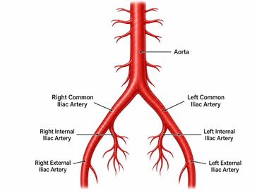
The aorta and iliac arteries are major blood vessels responsible for carrying oxygen-rich blood from the heart to the abdomen, pelvis, and lower extremities. Conditions affecting these arteries may include plaque buildup (atherosclerosis), narrowing, blockage, aneurysms, or reduced blood flow that can lead to symptoms such as leg pain, cr
The aorta and iliac arteries are major blood vessels responsible for carrying oxygen-rich blood from the heart to the abdomen, pelvis, and lower extremities. Conditions affecting these arteries may include plaque buildup (atherosclerosis), narrowing, blockage, aneurysms, or reduced blood flow that can lead to symptoms such as leg pain, cramping, weakness, poor circulation, or abdominal and pelvic discomfort. An aorta/iliac arterial duplex ultrasound is a specialized, non-invasive vascular study used to evaluate blood flow within these arteries and assess for abnormalities including stenosis (narrowing), occlusions, aneurysms, or plaque formation. Ultrasound imaging provides valuable information regarding circulation and arterial health, helping with early detection, monitoring, and guidance for appropriate treatment recommendations.
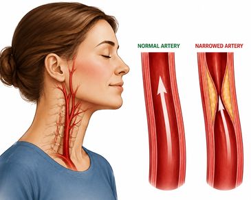
The carotid arteries are major blood vessels located in the neck that supply oxygen-rich blood to the brain. Over time, plaque buildup (atherosclerosis) can cause narrowing or blockage within these arteries, increasing the risk of stroke or transient ischemic attacks (TIA). A carotid artery duplex ultrasound is a specialized, non-invasive
The carotid arteries are major blood vessels located in the neck that supply oxygen-rich blood to the brain. Over time, plaque buildup (atherosclerosis) can cause narrowing or blockage within these arteries, increasing the risk of stroke or transient ischemic attacks (TIA). A carotid artery duplex ultrasound is a specialized, non-invasive vascular study used to evaluate blood flow within the carotid arteries and assess for plaque, narrowing (stenosis), or reduced circulation to the brain. This examination provides important information regarding stroke risk and overall vascular health. Early detection through ultrasound can help guide appropriate monitoring, medical management, or treatment recommendations to reduce the risk of serious neurological complications.

At EVS Imaging, we focus exclusively on vascular studies- delivering accurate comprehensive ultrasounds that support confident clinical decisions and better patient outcomes.
Our state-of-the-art imaging technology allows us to detect and diagnose medical conditions with unparalleled accuracy and speed. Our experienced team uses the latest techniques to provide accurate diagnoses.
Our expert ultrasound professionals are Registered Vascular Sonographers highly trained in performing vascular imaging. They provide precise, non-invasive studies to evaluate blood flow in the arteries and veins, assisting physicians in accurately diagnosing their patients conditions. Vascular ultrasound is not just something we do- it's our specialty.
We make vascular testing accurate, comfortable, and hassle-free. Our precise, non-invasive imaging services deliver fast, reliable results- because we believe in quality over volume, not rushed exams. Each study is performed thoroughly to provide clinically meaningful information that helps patients and providers move forward with the right treatment plan without unnecessary delays.
Our team of caring and compassionate professionals are here to support patients and physicians every step of the way. We offer personalized care and attention to ensure that everyone receives the best possible experience.